24-year-old male patient; with a history of falling to the floor two weeks ago; indicates severe pain in the jaw and severe limitation of jaw opening.
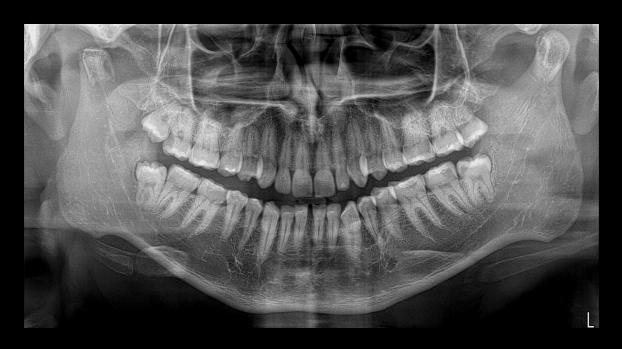
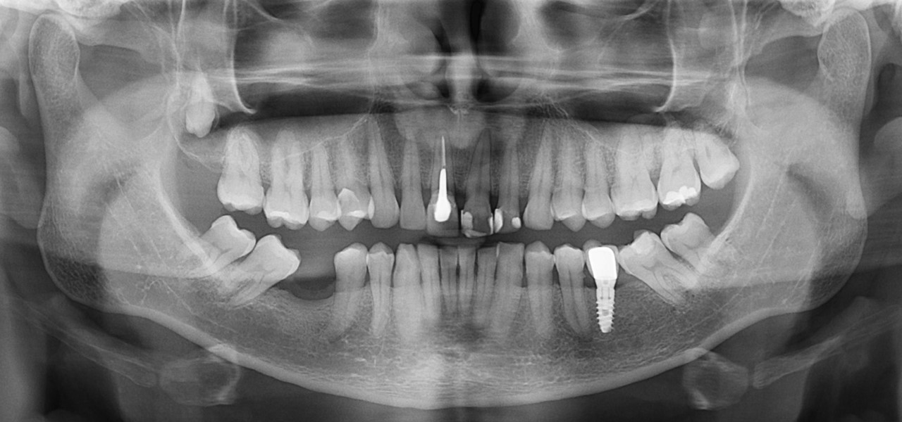
Figure 1
In the panoramic radiograph (Figure 1), a fracture is observed at the level of both mandibular condyles with displacement of fragments; Likewise, a trace of a mandibular fracture can be seen at the anterior level (between pieces 31 and 41) that extends to the mandibular base.
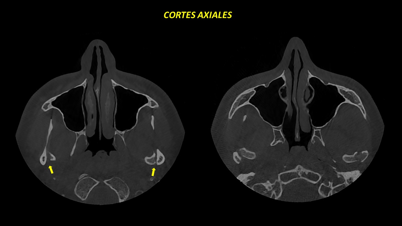
Figure 2
Figure 3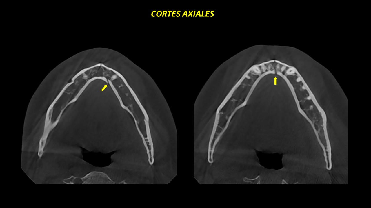
Figure 3
The evaluation of the volumetric tomography in axial sections (Figure 2 and 3) corroborates the fracture line of both mandibular condyles in an antero-posterior direction with anterior and medial displacement of the bone fragments. Furthermore, it is observed
the mandibular fracture at the level of the symphysis with extension from the top of the ridge to the base of the mandible.

Figure 4
In the coronal sections (Figure 4), an oblique fracture of both mandibular condyles is seen that extends from the superior articular surface to the internal subcondylar region; The medial and caudal displacement of the bone fragments is corroborated.
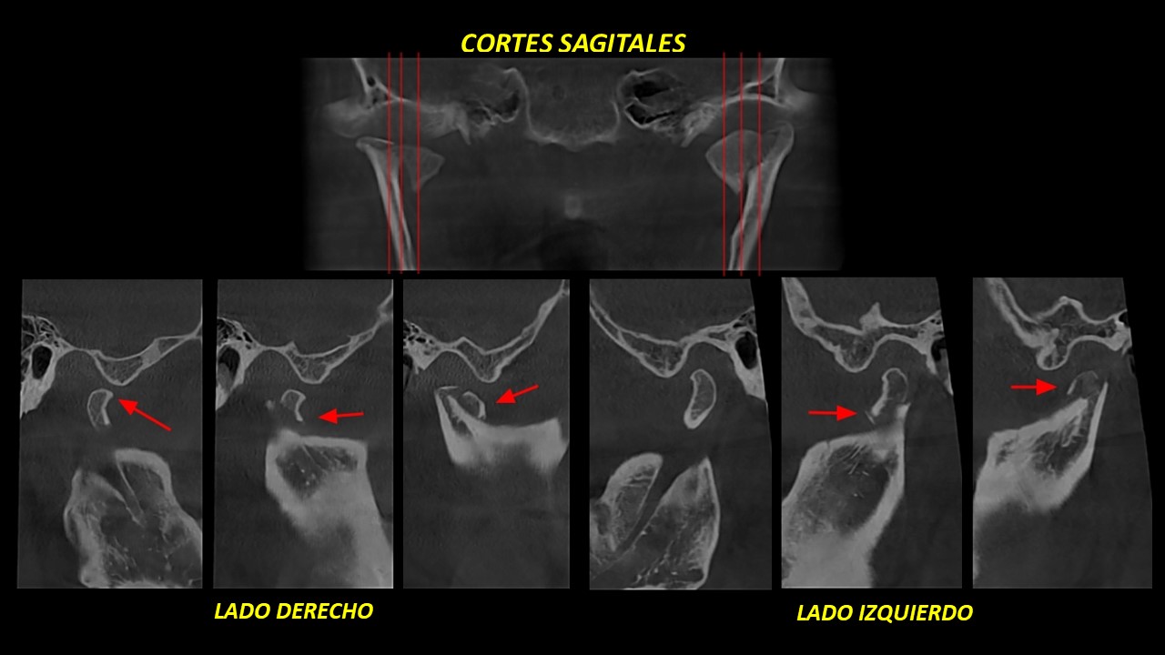
Figure 5
In the sagittal sections (Figure 5), anterior and caudal displacement of the condylar fragments can be seen; as well as distraction of the inter-articular spaces.
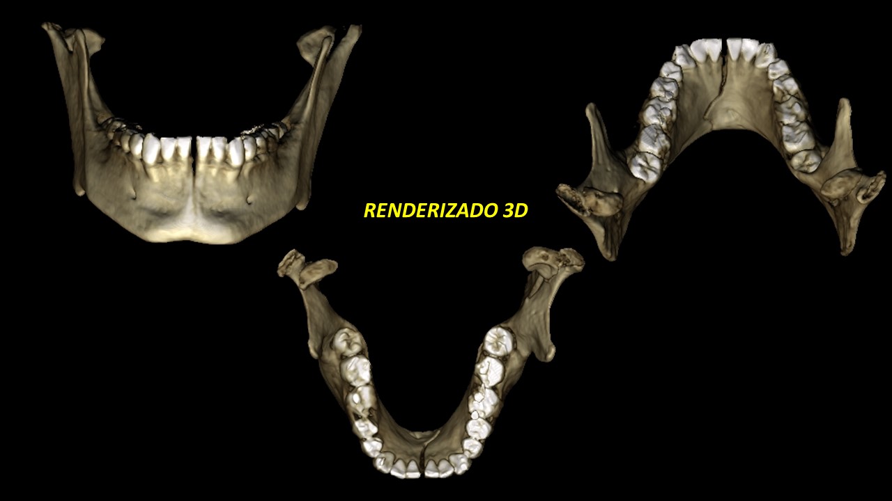
Figure 6
The 3D rendering (Figure 6) shows in a graphic and didactic way the bicondylar fracture with displacement of bone fragments; Likewise, the mandibular fracture at the level of the symphysis without displacement of fragments is corroborated.
Comments
The maxillofacial region is one of the most traumatized areas in the human body; Of all of them, condylar fractures represent between 18 and 24% of all jaw fractures.
The objective of imaging examination is to depict the presence, location, extent of fractures, displacement of fragments and foreign objects as a result of trauma. One or more combinations of 2D examinations can initially assist in an initial assessment of the severity of trauma at the bone level; However, three-dimensional 3D examinations such as computed tomography (CT) are ideal for the exact visualization of alterations; Of all of them, Cone Beam volumetric tomography (CBCT) may be the one of choice due to its simple technical design, low cost and reduced space requirements, facilitating dental professionals’ access to cross-sectional information from images with high spatial resolution; All this with the aim of making the most accurate diagnoses since the appropriate treatment will depend on it, which consists of stabilizing the bite in the initial occlusion and minimizing the risks of temporo-mandibular ankylosis.

Author: Dr. LUIS CUEVA
-
-
-
-
-
-
-
-
-
-
-
- Specialist in Oral and Maxillofacial Radiology at the University of San Martin de Porres (USMP).
- Master in Stomatology from the Scientific University of the South (UCSUR).
- Professor of Oral and Maxillofacial Radiology at the University of San Martin de Porres (USMP).
- Professor of Oral and Maxillofacial Radiology at the Peruvian University of Applied Sciences (UPC)
- Member of the International American Dental Maxillofacial Radiology (IADMFR).
- Member of the Latin American Association of Dentomaxillofacial Radiology and Imaging (ALARID).
- Oral and Maxillofacial Radiologist at Diagnostic Imaging Center (CDI).
-
-
-
-
-
-
-
-
-
-


